Clinical Example: Open Reduction Distal Phalanx Base Fractures with Wires and Pins
| Distal phalanx fractures are common and pose a variety of problems relating to the close proximity of nailbed, nerves, joint and tendon attachments and the fact that it represents a terminal vascular zone. These cases represent simple types of fixation which may be required for the small fragments of comminuted distal phalanx base fractures. |
| Click on each image for a larger picture |
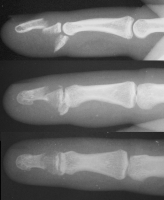
| Case 1. This patient sustained a closed comminuted fracture of the distal phalanx with dorsal-palmar split of the base and a completely displaced transverse fracture of the diaphysis. |


| A midline longitudinal palmar approach was used. This view shows a transverse pin across the dorsal proximal fragment, to be used as a path for interosseous wiring. Additional pins have been placed across each single cortex for later advancement. |

| A wire was passed through the dorsal proximal pin track and then around the palmar pins. After reduction and tightening this wire, the pins were advanced to engage the dorsal cortex. |

| Wiring was used to offset the strong pull of the flexor insertion. |

| All pins were cut below the surface of the skin. |

| Inury. |

| One week postop. |

| Three months postop, after removal of prominent pins in the office. |

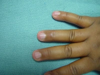
| Late appearance (ring finger) |

| Midline palmar scar. |

| Range of motion. |


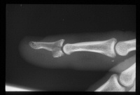
| Case 2. This patient sustained a closed displaced distal phalanx base fracture with dorsal-palmar split and dorsal subluxation of the larger fracture fragment. |


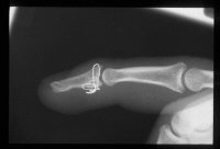
| This was managed with two wires - a cerclage wire, passed over the dorsum in a subperiosteal plane, and an interfragmentary palmar wire. The profundus tendon was found to have avulsed from the palmar fragment, and the tendon was reattached with sutures passed around the palmar component of the cerclage wire. A temporary longitudinal interfragmentary pin was also used (not shown). |


|
Search
for... distal phalanx base fracture phalanx small bone fixation
|
Case Examples Index Page | e-Hand home |